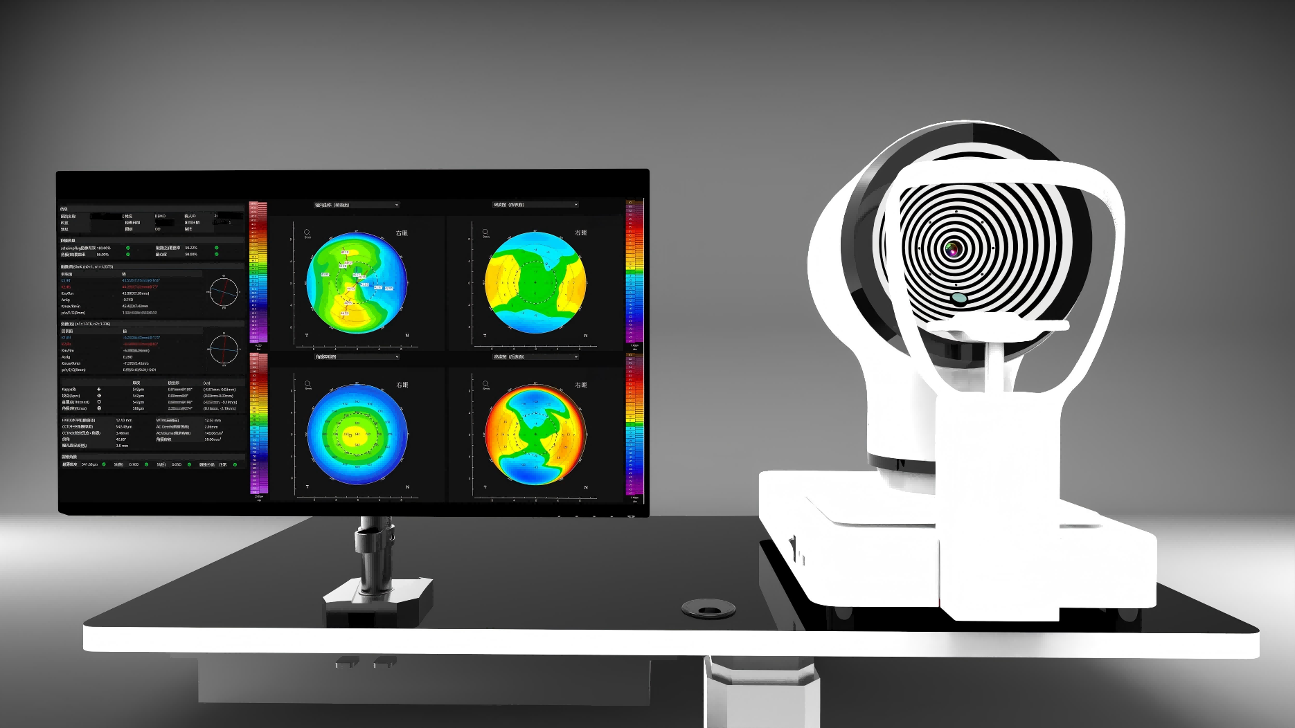
Professional Ophthalmic Imaging & Measurement
Ophthalmic Imaging & Measurement, Choose OptiV
12 professional ophthalmic imaging and measurement devices, trusted by 200+ ophthalmologists worldwide. Micron-level 3D reconstruction technology transforms anterior segment into quantifiable data for precise surgical planning beyond human vision limits.
2x Diagnostic Efficiency
NMPA Certified
Expert Technical Support

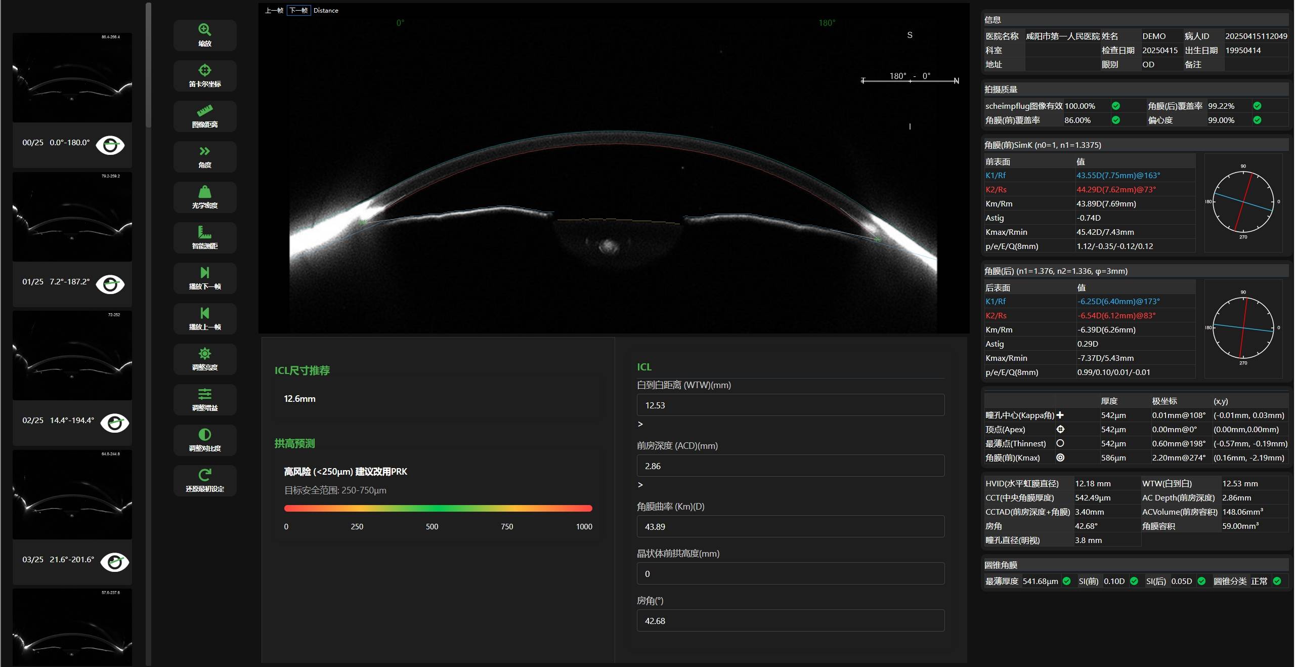
Ultra-wide Measurement
25mm scleral range, beyond traditional limits
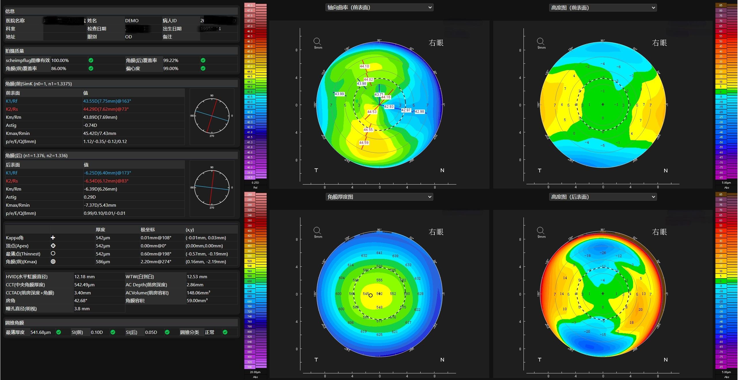
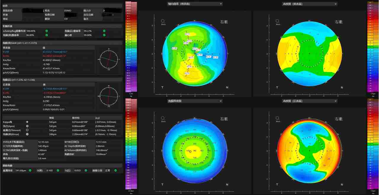
Depth Tomography
7mm anterior chamber depth analysis
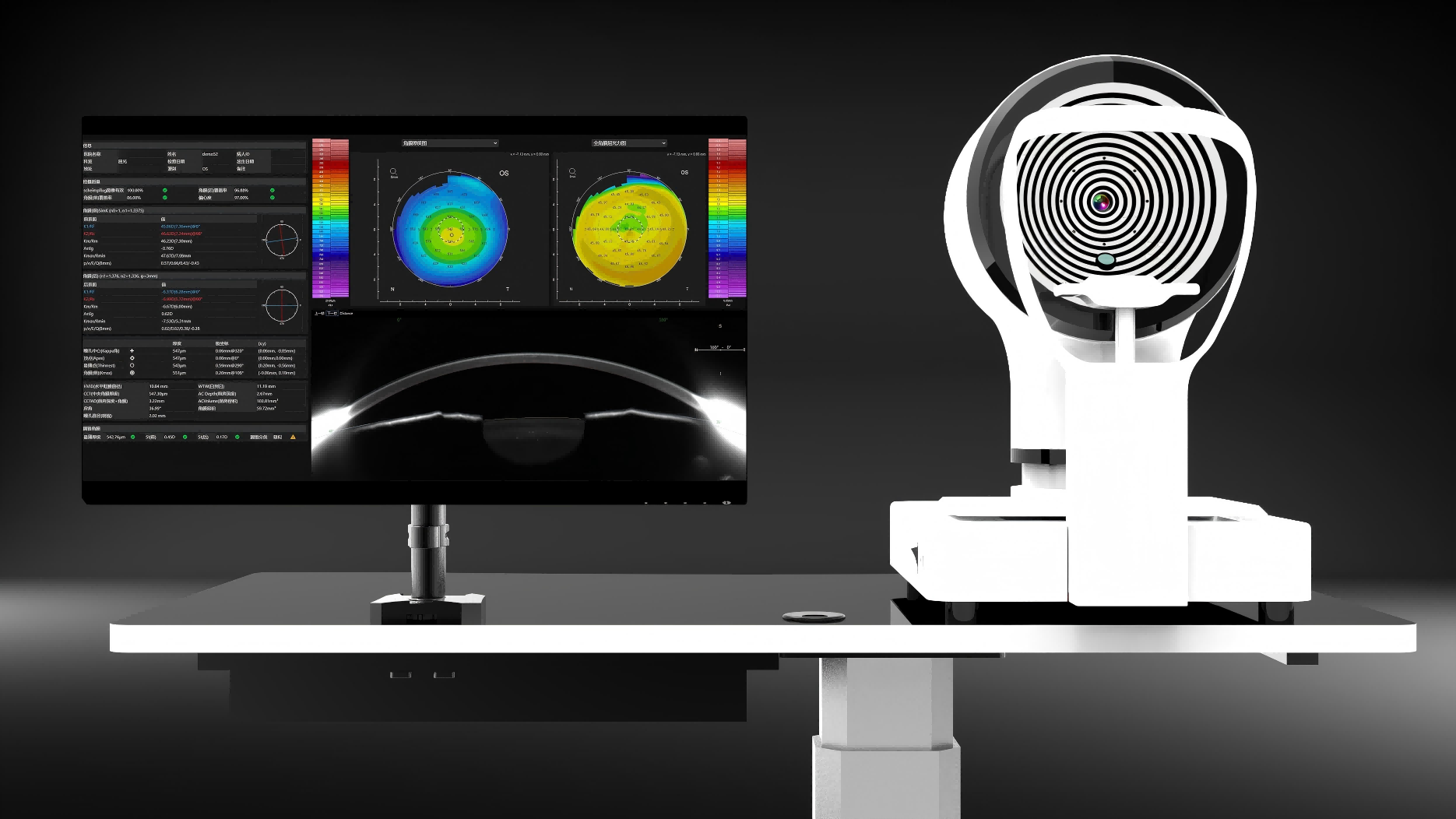
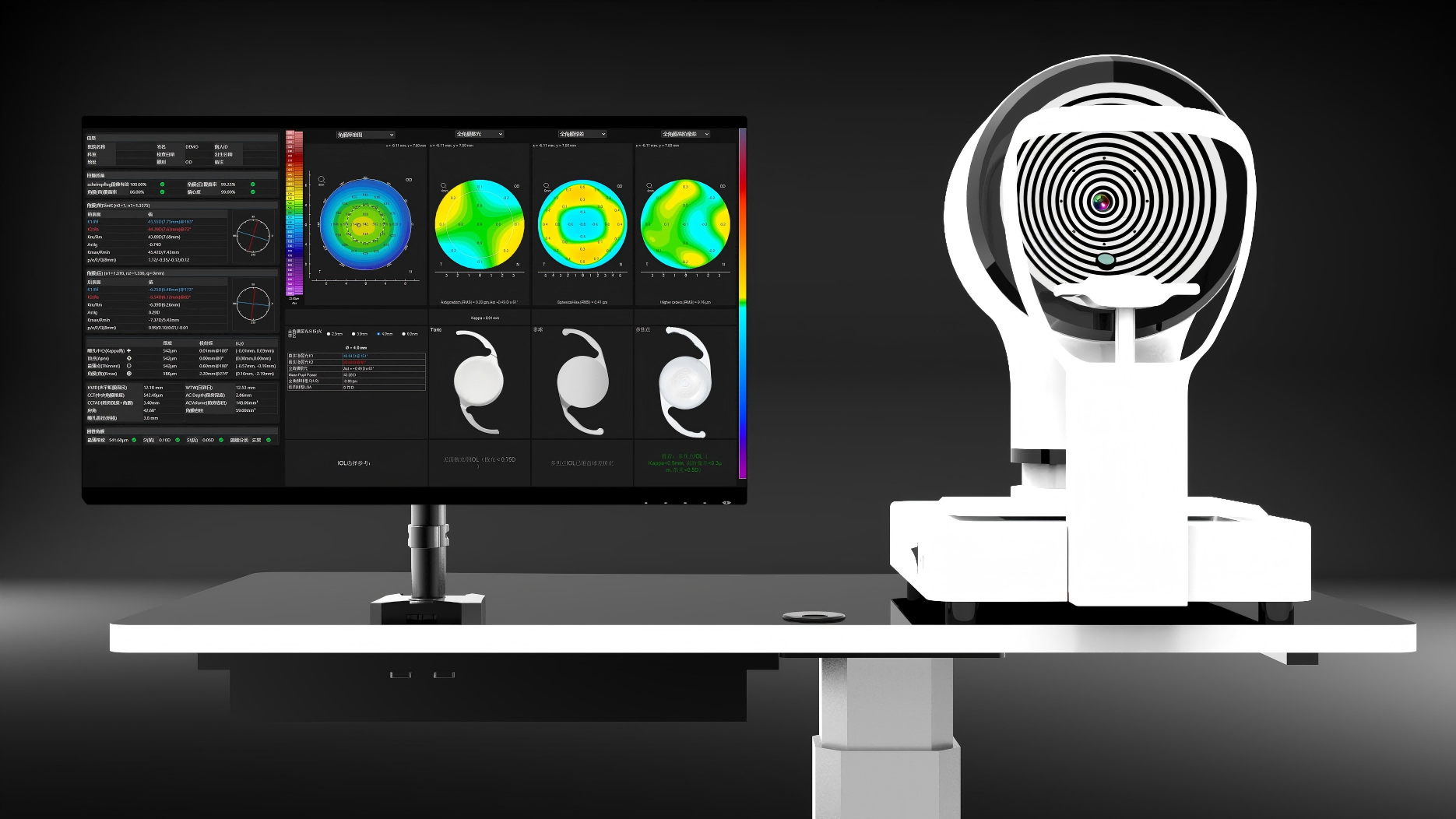
Dual Gold Standard
Placido disk + Scheimpflug fusion
200+
Partner Hospitals
99.9%
Measurement Accuracy
24/7
Tech Support